NCPV in Research 2020
Established in 1999, The National Collection of Pathogenic Viruses (NCPV) collects, conserves and distributes clinically relevant viruses in order to aid researchers worldwide (1). The collection is made up of over 300 viruses including many lesser-known viruses which have the potential to cause human disease (2). NCPV viruses are used within research both as the subject of investigation and also as positive controls. This article highlights several papers published in 2020 which include the use of NCPV strains.
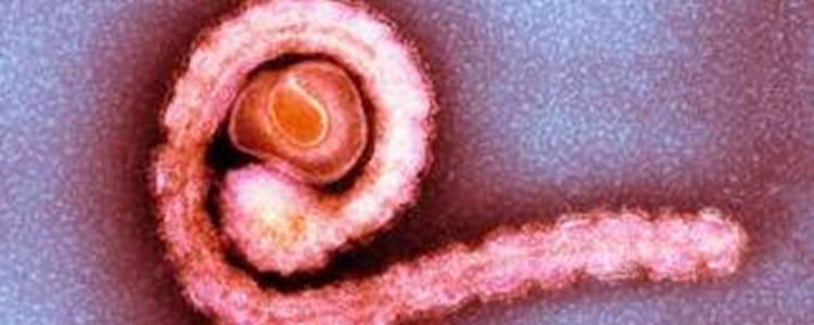
In recent years, there have been increased numbers of vector-borne viral outbreaks within Europe to naïve populations (3, 4). Some notable examples of these zoonotic viruses which cause disease include Ebola virus, West Nile virus and Zika virus (4). Increased concerns over future mosquito-borne viral disease outbreaks within Europe due to enhanced globalisation and climate change has amplified the necessity to conduct research in these regions. Chapman et al., (2020) investigated the laboratory potential of British mosquitos, such as Ochlerotatus detritus and Culex pipiens, to act as vectors for zoonotic equine arboviruses; including Venezuelan equine encephalitis virus P676 (0605153v) and Japanese encephalitis virus (JEV) strain CNS138-11 (0005281v) from NCPV’s collection. Their results demonstrate that British mosquitos can expectorate viral RNA at 7 days post-infection at a temperature between 21 and 24 °C, while after 21 days at 18 °C, 72% of Cx. pipiens produced JEV RNA within their saliva (3).
NCPV strains can also be utilised to study the intracellular sites where viral activities such as replication occur, as well as ways to inhibit viral processes (4, 5). While some viruses have had their intracellular processes precisely mapped, for example the replication factory of Bunyamwera virus, others have not been defined (5). In 2020, Davies et al., identified and characterised the cellular sites of Tula virus (0504102v) replication during early, peak and persistent phases of infection. Using immunofluorescent microscopy and transmission electron microscopy, it was observed that the Tula virus nucleocapsid protein was distributed within punctate and filamentous structures that increasingly co-localised within the Golgi apparatus, over the infection time course (5). This showed that Tula virus induced dramatic redistribution of the intracellular organelles enabling the establishment of a re-modelled Golgi apparatus for RNA synthesis (5). Research into the identification of where RNA synthesis occurs within the cell is important for treatment development.
Research has also been conducted into supporting outbreak responses and the reduction of risk in front-line diagnostics (4). One study investigated the use of X-ray irradiation which it was believed had the potential to allow diagnostic work at lower containment levels (4). Chemical and thermal methodologies for virus inactivation are already established, however, they have detrimental effects on the biological integrity of the specimens (4). Afrough et al., (2020) used Zika virus African strain MP1751 (1308258v) from NCPV, as one of the subjects of their study into the effect of X-ray irradiation as a method of virus inactivation (4). They discovered that soft X-rays (≤~20 keV), X-rays which can be filtered out via the use of 0.3 mm Cu filtration, are more effective at inactivation than hard X-rays (4). This research concluded that X-ray irradiation can be utilised for the successful inactivation of Zika virus (amongst others), under ambient conditions without loss of biological characteristics (4).
We would like to hear from you if you have used NCPV viruses in your research. Viruses should be cited in publications as: ‘Virus Name’ (NCPV ‘Catalogue Number’), and you could feature in one of our newsletters or on the Culture Collections website.
Culture Collections specialise in preserving and distributing viral strains. We offer a catalogue deposit service where your deposited organisms will be maintained by experienced staff, included in our online catalogue and made available to researchers around the globe.
Please click here if you would like to browse the NCPV catalogue and see all of our newest accessions.
References
- Nissimov, J., Campbell, C., Probert, I. and Wilson, W., 2020. Aquatic virus culture collection: an absent (but necessary) safety net for environmental microbiologists. Applied Phycology.
- Atkinson, B. 2020. From obscurity to notoriety - the evolving nature of viral diseases [Available online] www.phe-culturecollections.org.uk/news/ncpv-news/from-obscurity-to-notoriety-the-evolving-nature-of-viral-diseases (Accessed 20.01.2021)
- Chapman, G., Sherlock, K., Hesson, J., Blagrove, M., Lycett, G., Archer, D., Solomon, T. and Baylis, M., 2020. Laboratory transmission potential of British mosquitoes for equine arboviruses. Parasites & Vectors, 13(1).
- Afrough, B., Eakins, J., Durley-White, S., Dowall, S., Findlay-Wilson, S., Graham, V., Lewandowski, K., Carter, D. and Hewson, R., 2020. X-ray inactivation of RNA viruses without loss of biological characteristics. Scientific Reports, 10(1).
- Davies, K., Chadwick, B., Hewson, R., Fontana, J., Mankouri, J. and Barr, J., 2020. The RNA Replication Site of Tula Orthohantavirus Resides within a Remodelled Golgi Network. Cells, 9(7).
Prepared by Callum Robertson