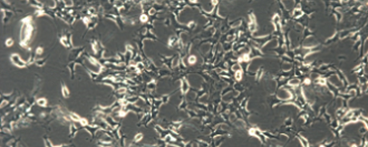
Sticky Issues with 293 Cells
Often with years of experience of growing many different cell types such as cancer derived cells, fibroblast cell lines or primary cells, cell culturists pride themselves on their ability to maintain sterility and sustain growth of cell cultures to generate reproducible and consistent results. The impact of failure, either through microbial contamination, cell death or the inability for cells to attach and grow can be devastating to morale and impact on study costs and time-lines. These frustrations can be particularly acute when they occur with a commonly used cell line such as 293 (ECACC 85120602) (also known as ‘HEK 293’).
The 293 cell line was developed in 1977 by transformation of human embryonic kidney cells with adenovirus 5 DNA1 and since inception it has become the second most commonly used cell line in laboratories world-wide2 after HeLa. Amongst many other applications 293 cells are used in the drug discovery field for efficacy testing, as a transfection host and in virology. The Cellosaurus database3 shows that there are currently over 90 different variant cell lines based on 293 and literature search reveals there are thousands of publications that reference the cell line. 293 cells are commonly found and grown in many tissue culture facilities as strains that grow as both adherent and non-adherent phenotypes. However, most of the strains are described as adherent.
There is an expectation when working with adherent cell lines that they will attach to their substrate within a few hours of resuscitation or subculture; they will remain attached between subcultures and to micro-titre plates for the duration of an assay. Loss of cellular adherence may give the false impression that cultures have died or have been subject to microbial contamination during routine culture. Loss of adherence can also be problematic during cell based assays. Often when a researcher encounters problems of adherence or apparent low viability they tend to look for shortcomings in their technique or a fault in the batch of cells used rather than looking for an innate biological cause or a fundamental problem with the procedure they are following.
Although there is a lack of peered reviewed literature regarding problems with attachment of 293 cells, it is generally agreed that the cell line is semi-or loosely adherent. From experience in ECACC, unlike many other cell types, 293 cells can take several days to attach following resuscitation from frozen and it is important to have patience at this critical stage. There is a wealth of anecdotal evidence of issues regarding 293 cell attachment issues in on-line discussion platforms such as ‘ResearchGate’. Switching of plastic-ware manufacturers or the use of collagen or Poly-D-Lysine (PDL) coated plates and flasks are often suggested as solutions.
There may, however, be innate explanations to the issues experienced with 293 cells which illustrate the need for knowledge of the biology of cell lines and the adaptation of techniques to accommodate specific cellular requirements.
Attachment of cells to each other and to their substrate is a fundamental property of multicellular organisms and key to the development and cellular organisation in organs and tissues. The mechano-sensing pathways that control this process, such as the Hippo pathway, are highly conserved throughout the animal kingdom. Attachment and spreading of cells is a complex biological process that is primarily mediated and controlled by actin microfilaments of the cell cytoskeleton. Actin filaments are not simply a rigid scaffold within the cell but the core of a dynamic system that involves:
- Polymerisation, depolymerisation and branching of actin.
- Close interaction and control of the extracellular matrix (ECM) (e.g. fibronectin).
- Involvement of attachment and molecular mechanical sensing through integrins (for cell to ECM attachment) and cadherins (for cell to cell attachment).
Not only does this complex interconnected system allow directed cellular movement and attachment it also triggers cytoplasmic signalling pathways through kinases, phosphatases and small GTPases to affect a wide range of cellular processes such as proliferation, survival and gene expression. It is likely that defects in actin expression or processes that interfere with polymerisation will have a marked effect on cellular adhesion4.
293 cells appear to have a distinctly different actin cytoskeleton to other commonly used cell lines such as cancer cell lines and fibroblasts as was demonstrated by Haghparast et al5 to the extent that these authors recommend that 293 cells should be considered as having a specific actin cytoskeleton type as distinguished by ‘immature’ actin.
How the unique actin cytoskeleton observed in 293 cells arose, could in part be explained by the original generation of the cell line using sheared adenovirus 5 DNA fragments. Viral pathogens are known to induce actin polymerisation in host cells to enable infection and replication. Nuclear actin is known to play a role in adenovirus replication and infection6 resulting in reorganisation of the host cytoskeleton7. During viral infection adenovirus 5 is trafficked to the nucleus via the actin cytoskeleton and it is postulated that the irregularly shaped nucleus observed in 293 cells may be a residual effect on the cytoskeleton resulting from the cell line’s original treatment with viral DNA. It has been observed that the pattern of adenovirus trafficking in subsequent infections with the virus is different in 293 cells than observed in either HeLa or A549 cancer cells8 indicating that 293 cells may be ‘primed’ for infection.
To add to this supposition, 293T (ECACC 12022001) (often mistakenly used synonymously with 293) is a commonly used cell line derived from parental 293 cells through transfection with the temperature sensitive neomycin / G418 resistant tsA1609 allele of SV40 large T antigen9. SV40 virus is also known to have disruptive effect on cellular actin10.
Reproducible and reliable cell culture is generally achieved through optimal cell culture techniques. Protocols will often dictate that cell culture media and reagents are warmed prior to use and that cells are maintained at a constant 37°C. In reality, however, these constraints are often relaxed, sometimes through time-pressures and conditions in the laboratory (such as allowing reagents to cool during cell culture, or not checking they have adequately warmed before use). There may also be unavoidable cooling of flasks or assay plates to a temperature below 37°C when they are transferred to microscopes or plate readers for examination and analysis. For many common cell lines, the use of room temperature conditions or cool media do not have significant effects on cell adherence. Tight temperature control of 293 cells however, is critical. Although it has been shown that the optimum temperature range for protein expression in 293 cells may be in the 33-35°C range, it is apparent that cellular attachment is also temperature dependent. Reducing the temperature of 293 cultures to 30°C can result in up to 60% loss of the cells from the monolayer11. Temperatures of less than 30°C should be avoided and it is imperative to use pre-warmed medium and reagents when working with the cells. If 293 cells are observed to detach from a substrate, this may not indicate a problem with viability, it may simply be the case that at some point the culture might have cooled below 30°C for a short while. This issue may become particularly apparent if growing cultures are transferred from one laboratory to another (for example in the case of a ‘Growing Order’ of cells from ECACC). If the cells become detached it is important to check cellular viability, and as with resuscitation, to be patient and prepared to wait for several days for the cells to re-attach. If the cells are required for cell based assay running at a temperature less than 30°C it is recommended that the assay protocol is optimised to a time-window that allows data capture before cellular detachment or coatings such as Poly-D-Lysine (PDL), collagen or optimised treated plastics such as CellBind™ (Corning®) are used.
The final consideration with working with 293 and its derivatives is that the cell line is reported as being inherently genetically unstable with a hypertriploid karyotype12. There is therefore a need to be particularly attentive to avoid exerting the cells to selective pressures that might induce phenotypic or genotypic change. Uncontrolled switching of cell culture medium; allowing cells to become overconfluent; not being disciplined in maintaining strict sub-culture regimes or simply keeping cells in culture for extended periods may induce unwanted and irreparable damage to any cell line. 293 and its derivatives, however are particularly susceptible to genotypic drift induced by external factors. This is due to an inherent defect in the cell line’s DNA mismatch repair mechanism13. This factor goes some way to explain how dozens of different 293 derived cell lines each with subtly different genotypes, Short Tandem Repeat (STR) profiles and karyotypes14 have arisen since the cell line was first generated. The control of passage numbers used for experiments and assays using controlled Master and Working Banking procedures15 is therefore imperative with 293 cells.
Summary
It should not be assumed that a “generic” cell culture protocol that is suitable for continuous cancer derived cell lines will be suitable for 293 cells.
The cytoskeleton of the 293 cell line and its derivatives differs from those found in cancer derived cell lines or in fibroblast cell lines. These differences, apparent in the unique nature of actin microfilaments, may help explain observed issues regarding cell attachment.
It may be necessary to evaluate cell culture plastics from different suppliers to ensure 293 cell attachment in specific processes such as cell based assays. It could also be useful to explore the use of coatings such as Poly-D-Lysine (PDL), collagen or CellBind™ to increase cellular attachment.
On resuscitation from frozen, 293 cells will take longer to attach than other cell lines and patience is required as attachment may take several days.
293 cells will detach if their temperature drops below 30°C. Keeping cell culture reagents warmed and avoiding drops in temperature are essential with 293 cells. If cells detach, do not assume the culture has died. Sample the culture and test for cell viability. The cells may re-attach after culture at 37°C, however, this could take several days.
293 cells are genetically unstable and more prone to genotypic drift than many other cell lines due to a defective DNA mismatch repair mechanism. Avoid exposing the cells to unnecessary selection pressures such as inconsistent subculture conditions, uncontrolled changes in medium composition, allowing cells to become over-confluent or prolonged periods in culture. Genotypic and phenotypic drift may become apparent through behavioural changes in the cells. Well-maintained cell banks should be established and passage numbers controlled.
It is important to regularly test human cell cultures by STR profiling as assurance against cross contamination or misidentification. STR profiling of 293 cells has an added benefit as changes in STR profile may alert genotypic drift in the cells.
References
1. Graham, F. & Smiley, J. Characteristics of a Human Cell Line Transformed by DNA from Human Adenovirus. J. Gen. Virol.36, 59–74 (1977).
2. Lin, Y.-C. et al. Genome dynamics of the human embryonic kidney 293 lineage in response to cell biology manipulations. Nat. Commun.5, 1–12 (2014).
3. Bairoch, A. The Cellosaurus, a Cell-Line Knowledge Resource. J. Biomol. Tech. JBT29, 25–38 (2018).
4. Bachir, A. I., Horwitz, A. R., Nelson, W. J. & Bianchini, J. M. Actin-Based Adhesion Modules Mediate Cell Interactions with the Extracellular Matrix and Neighboring Cells. Cold Spring Harb. Perspect. Biol.9, (2017).
5. Haghparast, S. M. A., Kihara, T. & Miyake, J. Distinct mechanical behavior of HEK293 cells in adherent and suspended states. PeerJ3, (2015).
6. Fuchsova, B., Serebryannyy, L. A. & de Lanerolle, P. Nuclear Actin and Myosins in Adenovirus Infection. Exp. Cell Res.338, 170–182 (2015).
7. Staufenbiel, M., Epple, P. & Deppert, W. Progressive reorganization of the host cell cytoskeleton during adenovirus infection. J. Virol.60, 1186–1191 (1986).
8. Yea, C., Dembowy, J., Pacione, L. & Brown, M. Microtubule-Mediated and Microtubule-Independent Transport of Adenovirus Type 5 in HEK293 Cells. J. Virol.81, 6899–6908 (2007).
9. DuBridge, R. B. et al. Analysis of mutation in human cells by using an Epstein-Barr virus shuttle system. Mol. Cell. Biol.7, 379–387 (1987).
10. Graessmann, A., Graessmann, M., Tjian, R. & Topp, W. C. Simian virus 40 small-t protein is required for loss of actin cable networks in rat cells. J. Virol.33, 1182–1191 (1980).
11. Lin, C.-Y. et al. Enhancing Protein Expression in HEK-293 Cells by Lowering Culture Temperature. PLoS ONE10, (2015).
12. Stepanenko, A. A. & Dmitrenko, V. V. HEK293 in cell biology and cancer research: phenotype, karyotype, tumorigenicity, and stress-induced genome-phenotype evolution. Gene569, 182–190 (2015).
13. Panigrahi, G. B., Slean, M. M., Simard, J. P. & Pearson, C. E. Human Mismatch Repair Protein hMutLα Is Required to Repair Short Slipped-DNAs of Trinucleotide Repeats. J. Biol. Chem.287, 41844–41850 (2012).
14. Binz, R. L. et al. Identification of novel breakpoints for locus- and region-specific translocations in 293 cells by molecular cytogenetics before and after irradiation. Sci. Rep.9, (2019).
15. Geraghty, R. J. et al. Guidelines for the use of cell lines in biomedical research. Br. J. Cancer111, 1021–1046 (2014).
Written by Jim Cooper, January 2021